
Patient History
67 year old female with Child-Pugh B8 cirrhosis who presents with an isolated 4 cm segment 4 HCC. The patient is a non-operative candidate due to comorbidities and the extent of liver disease and was referred to VIR for liver-directed therapy.
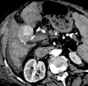
Pre-Procedure CTA
The pre-procedure CTA demonstrated a single hyper vascular mass within segment 4 supplied by an apparent single feeding branch of the right hepatic artery.
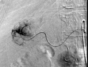
Mapping Angiogram
The mapping angiogram confirmed the presence of tumor hypervascularity arising from a small branch of the right hepatic artery.
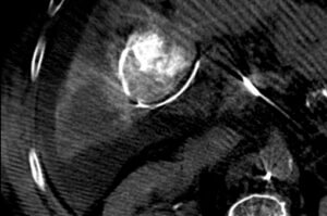
Cone-beam CT
A cone-beam CT was performed during the mapping angiogram to better characterize the vascular anatomy of the tumor and identify any non-target enhancement of normal liver. A single feeding artery to the tumor was confirmed. This artery did not contribute blood supply to normal liver. On the treatment day, the tumor was treated via superselective administration of y-90 glass microspheres in a radiation segmentectomy approach.
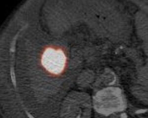
Post-y90 PET-CT
Following y-90 administration, the patient was transferred to the PET scanner for a post-infusion PET-CT, which confirmed complete tumor coverage.